
Key facts
- Glaucoma is a chronic optic-nerve disease, the “silent thief of sight”: painless and symptom-free for years in its open-angle form (90 % of cases).
- It cannot be cured but is stabilised effectively: eye drops, SLT laser or surgery — lifelong treatment with regular monitoring.
- Screening is recommended from age 40, from 30 with risk factors; the risk is multiplied by 4–9 if a first-degree relative is affected.
- Lost nerve fibres do not regenerate: early detection is what preserves vision.
The silent thief of sight
Glaucoma is a chronic, progressive disease of the optic nerve, most often linked to raised intraocular pressure (IOP). It gradually destroys the nerve fibres that carry images to the brain — without pain or noticeable symptoms for years. Worldwide it is the second cause of blindness after cataract, affecting around 80 million people; in France more than a million people have glaucoma and nearly half don’t know it.

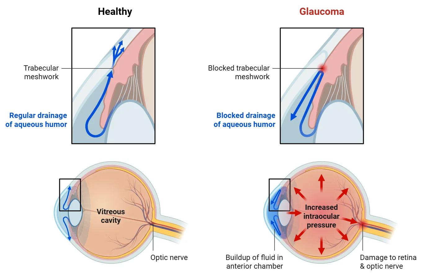
The eye continuously produces a clear fluid (aqueous humour) that drains through a microscopic filter, the trabecular meshwork. When drainage is impaired, pressure rises (normal range: 10–21 mmHg, with physiological daily variations of up to 5 mmHg) and slowly compresses the optic nerve fibres. Vision loss starts in the periphery, sparing central vision for a long time — which is exactly why the diagnosis is so often late.

Risk factors
Non-modifiable
- Age over 40 — risk doubles every decade
- Family history — risk ×4 to ×9 with an affected first-degree relative
- African or Caribbean ancestry — prevalence 4–5× higher
- High myopia (beyond −6 D)
- Thin cornea (under 520 µm)
Modifiable / associated
- Raised intraocular pressure — the main treatable factor
- Long-term corticosteroid use (drops or systemic)
- Diabetes, high blood pressure, sleep apnoea
- Pseudoexfoliation syndrome
The main types
Open-angle glaucoma (90 %)
The drainage angle stays open but the meshwork clogs progressively. Slow, totally asymptomatic evolution over years — detected only by screening. Peripheral field loss goes unnoticed until late.
Acute angle-closure glaucoma — emergency
The iris suddenly blocks drainage: pressure soars above 40 mmHg with severe eye pain, red eye, blurred vision, coloured halos, nausea. This is an immediate emergency — call us or go to a 24/7 eye emergency department. A preventive YAG laser iridotomy protects predisposed (narrow-angle) eyes.
Normal-tension glaucoma
15–25 % of open-angle cases: the nerve deteriorates despite pressure within the normal range, probably through vascular fragility of the optic nerve (associated with migraine, Raynaud’s phenomenon, nocturnal low blood pressure). A “normal” pressure reading alone can never rule out glaucoma — only a complete work-up can.
Pigmentary glaucoma
Pigment granules released from the back of the iris clog the drainage meshwork. Typically affects young myopic men (20–45), with possible pressure spikes after physical exercise.
Pseudoexfoliative glaucoma
An abnormal fibrillar protein accumulates in the drainage angle, often asymmetrically between the two eyes, in patients over 60. Multiplies the glaucoma risk by 5–10 and sometimes responds less well to drops, requiring earlier laser or surgery.
Other secondary glaucomas
Steroid-induced (5–6 % of people respond to prolonged corticosteroids with a pressure rise — monitored in every long-term steroid user), post-traumatic (even years after an eye injury) and neovascular (a complication of diabetic retinopathy or vein occlusion, requiring urgent combined treatment).
The glaucoma work-up at IPO
Intraocular pressure & corneal thickness
Pressure measured by air-puff or Goldmann applanation tonometry, always interpreted against corneal thickness (pachymetry): a thin cornea under-estimates the true pressure.
OCT of the optic nerve
The Zeiss Cirrus OCT maps the retinal nerve fibre layer and ganglion cell complex, compared to an age-adjusted normative database. It detects fibre loss before any visual field defect appears — the key to early diagnosis.
Gonioscopy
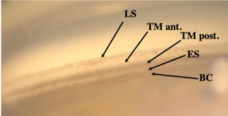
Direct examination of the drainage angle with a contact lens, to classify the glaucoma (open or narrow angle) and guide treatment.
Humphrey visual field
Automated perimetry quantifies any functional loss and provides the baseline for lifelong monitoring of progression.

Treatments
Eye drops
First-line treatment: daily pressure-lowering drops (prostaglandins, beta-blockers, and combinations). Effective, but require rigorous lifelong compliance.
SLT laser
Selective Laser Trabeculoplasty stimulates the drainage meshwork in a 5-minute painless clinic procedure. Can replace or defer drops; repeatable. Performed on site at IPO. For narrow angles, a preventive YAG iridotomy protects against acute angle-closure attacks.
Surgery
When drops and laser are insufficient: micro-invasive glaucoma surgery (MIGS — microscopic stents and bypasses, often combined with cataract surgery), deep sclerectomy or trabeculectomy, performed by our glaucoma surgeons in partner clinics.
Treatment does not restore lost fibres — it protects what remains. This is why regular follow-up (OCT + visual field, usually every 6–12 months) is as important as the treatment itself: comparing each examination to the previous ones is what detects progression early enough to step up treatment before vision is affected.
Frequently asked questions
Can glaucoma be cured?
No, but it can be stabilised very effectively. The goal of every treatment is to lower eye pressure enough to stop further nerve damage. Vision already lost cannot be restored — hence the importance of early screening.
I have no symptoms — why should I be screened?
Because open-angle glaucoma has none until late. From age 40 (30 with a family history or other risk factors), a screening visit with pressure measurement and OCT takes less than an hour and can save decades of sight.
Is the follow-up compatible with living abroad part of the year?
Yes. We coordinate with your home ophthalmologist, provide reports in English, and OCT/visual field data can be shared so monitoring stays continuous wherever you are.
Book a glaucoma screening
Pressure check, OCT of the optic nerve and specialist consultation — in English, all in one visit.