
Key facts
- Vitrectomy is the reference operation of the retina (epiretinal membrane, macular hole, retinal detachment), under local or general anaesthesia, painless.
- Duration: 30–45 minutes for a membrane or macular hole, 45–90 minutes for a standard detachment.
- Results: over 90 % of macular holes closed, 85–95 % of retinas reattached at the first operation.
- No flying and no driving while gas remains in the eye; the two eyes are never operated at the same time.
- IPO runs an on-call surgical service for retinal detachments.
The retina in brief
The retina is a neurosensory tissue about 0.5 mm thick lining the inside of the eye — a true extension of the brain that converts light into nerve signals. The macula (5.5 mm across) provides detailed central vision: reading, faces, colours. The peripheral retina handles night vision and the visual field — and is the zone most vulnerable to tears. This is why a peripheral tear or detachment can progress silently before central vision is hit.

Warning signs — when it’s urgent
See an ophthalmologist the same day if you notice:
- Sudden flashes of light (like lightning in the corner of the eye)
- A sudden shower of floaters (black dots, soot rain)
- A dark curtain or shadow spreading across your field of vision
- Sudden drop in vision in one eye
These can signal a retinal tear or the start of a retinal detachment — an absolute surgical emergency. Call +33 1 43 27 10 69: IPO sees eye emergencies the same day, Monday to Saturday, and runs an on-call surgical service for retinal detachments.
Conditions we operate
Retinal detachment
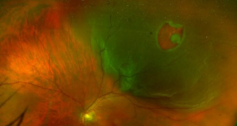
In the most common (rhegmatogenous) form, a tear lets fluid seep under the retina, peeling it off. Risk factors: high myopia, previous cataract surgery, trauma, age over 50. Treated by vitrectomy + laser + gas (or silicone oil) tamponade. Prognosis is best when the macula is still attached — hence the urgency. Rarer forms exist: tractional (fibrous membranes pulling on the retina, mainly in advanced diabetic retinopathy) and exudative (fluid accumulation from inflammatory or tumoural disease, usually treated medically).
Macular hole
A small opening at the very centre of the retina causing a central dark spot and distorted lines. Vitrectomy with internal limiting membrane peeling and a gas bubble closes over 90 % of holes.
Epiretinal membrane
A fine cellophane-like membrane grows on the macula, wrinkling it: distorted vision, progressive blur. Peeled surgically under the microscope when vision drops or distortion becomes disabling.
Vitreous haemorrhage & diabetic complications
Bleeding into the eye (diabetic retinopathy, retinal tear, vein occlusion) causing sudden vision loss. Vitrectomy clears the blood and treats the cause with endolaser.

A word about high myopia
A highly myopic eye (beyond −6 D) is longer, with a stretched, thinner retina exposed to peripheral tears, myopic foveoschisis, macular hole and retinal detachment. If you are highly myopic, regular dilated retinal examinations (with OCT and widefield imaging, both available at IPO) are the best protection — peripheral weak spots can be secured with preventive laser before they cause a detachment.
Vitrectomy: how it works
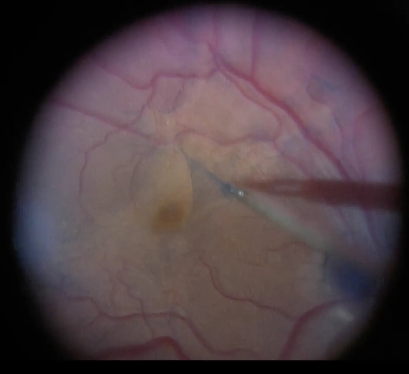
Vitrectomy removes the vitreous — the transparent gel filling 80 % of the eye — to access and treat the retina directly. Modern instruments are 0.4–0.6 mm in diameter (23, 25 or 27 “gauge”, chosen by the surgeon according to the complexity of the case), inserted through three self-sealing micro-incisions: no stitches in most cases, minimal inflammation and faster recovery with the finest calibres.
Anaesthesia
Local anaesthesia (an injection around the eye, not into it) in most cases, or general anaesthesia if preferred. The operation is painless.
Removal of the vitreous
A micro-guillotine cutter (up to 20,000 cuts/minute) removes the gel while an infusion line keeps the eye pressure stable.
Retinal repair
Depending on the condition: membrane peeling with micro-forceps after vital staining, laser around tears, drainage of sub-retinal fluid.
Tamponade
The eye is filled with a gas bubble (SF6 or C3F8, self-resorbing in 2–8 weeks) or silicone oil for complex cases, holding the retina in place while it heals.
After the operation — important for travellers
- No flying while gas remains in the eye. At altitude the bubble expands and can cause a dangerous pressure rise. Depending on the gas, this means 2 to 8 weeks without air travel — essential to plan if you live abroad. Your surgeon checks the bubble at each visit and tells you when flying is safe.
- No driving until the gas has resorbed and vision allows.
- Positioning: for macular holes, face-down positioning may be asked for a few days to keep the bubble pressing on the hole.
- Follow-up: day 1, week 1, then monthly until healing is complete — all at IPO.
- Vision recovers progressively over weeks to months depending on the condition and how early it was treated.
Frequently asked questions
Is vitrectomy painful?
No — it is performed under local or general anaesthesia. Mild foreign-body sensation and redness for a few days afterwards are normal.
How long before I recover vision?
While gas fills the eye, vision is very blurry — this is expected. As the bubble resorbs, vision clears progressively. Final recovery takes weeks to months and depends on the condition and how early surgery was done.
I'm only visiting Paris — can you operate my retinal detachment here?
A retinal detachment must be operated where you are — it cannot wait for a flight home (flying is dangerous with a fresh detachment and forbidden with intraocular gas). Our on-call retinal surgeons handle the emergency, then coordinate follow-up with your ophthalmologist at home, with reports in English.
Book a retina consultation
OCT imaging, widefield retinal photography and specialist retinal surgeons — same-day emergency slots Monday to Saturday.